
Mouse Secondary Antibodies


Biotium Endoglin / CD105 (Angiogenesis Marker)(ENG/3269), 0.2mg/mL
CD105/Endoglin is a Type I membrane glycoprotein located on cell surfaces and is part of the TGF-beta receptor complex. This protein has been found on endothelial cells, activated macrophages, fibroblasts, and smooth-muscle cells. Endoglin has a role in the development of the cardiovascular system and in vascular remodeling. Its expression is regulated during heart development. CD105 is highly expressed in endothelial cells during tumor angiogenesis and inflammation, with weak or negative expression in vascular endothelium of normal tissues. Angiogenesis is a promising prognostic marker in a variety of tumors. Endoglin is a more specific and sensitive marker for tumor angiogenesis than CD31 or CD34, as it labels only newly-formed blood vessels and may serve as a prognostic marker for prostate adenocarcinoma, and cancers of the lung, stomach, breast, and brain.Primary antibodies are available purified, or with a selection of fluorescent CF Dyes and other labels. CF Dyes offer

Biotium CD64(10.1), 1mg/mL
CD64 is an Fc receptor that plays a putative role in the initiation of cell-mediated cytotoxicity. Thus far, three genes encoding four distinct CD64 transcripts have been described. CD64 has been shown to associate with signal transducing subunit of the high affinity IgE receptor. Src family kinases Hck and Lyn show increased kinase activity and will co-immunoprecipitate with CD64 subsequent to receptor cross linking. CD64 is constitutively expressed on monocytes and macrophages; exposure of granulocytes to cytokines such as IFN-gamma and G-CSF can induce CD64 expression.Primary antibodies are available purified, or with a selection of fluorescent CF Dyes and other labels. CF Dyes offer exceptional brightness and photostability. Note: Conjugates of blue fluorescent dyes like CF405S and CF405M are not recommended for detecting low abundance targets, because blue dyes have lower fluorescence and can give higher non-specific background than other dye colors.
Biotium Adipophilin / Perilipin-2 (Marker of Lipid Accumulation) (ADFP/2755R), Biotin conjugate, 0.1mg/mL
Perilipin-2, also known as Adipophilin, belongs to the perilipin family, members of which coat intracellular lipid storage droplets. This 48 kDa protein is associated with the lipid globule surface membrane material, and maybe involved in development and maintenance of adipose tissue. However, it is not restricted to adipocytes as previously thought, but is found in a wide range of cultured cell lines, including fibroblasts, endothelial and epithelial cells, and tissues, such as lactating mammary gland, adrenal cortex, Sertoli and Leydig cells, and hepatocytes in alcoholic liver cirrhosis, suggesting that it may serve as a marker of lipid accumulation in diverse cell types and diseases.Primary antibodies are available purified, or with a selection of fluorescent CF Dyes and other labels. CF Dyes offer exceptional brightness and photostability. Note: Conjugates of blue fluorescent dyes like CF405S and CF405M are not recommended for detecting low abundance targets, because blue
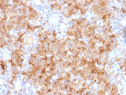
Biotium Chromogranin A / CHGA (Neuroendocrine Marker), Biotin conjugate, 0.1mg/mL
Chromogranin A is present in neuroendocrine cells throughout the body, including the neuroendocrine cells of the large and small intestine, adrenal medulla and pancreatic islets. It is an excellent marker for carcinoid tumors, pheochromocytomas, paragangliomas, and other neuroendocrine tumors. Co-expression of chromogranin A and neuron specific enolase (NSE) is common in neuroendocrine neoplasms. Reportedly, co-expression of certain keratins and chromogranin indicates neuroendocrine lineage. The presence of strong anti-chromogranin staining and absence of anti-keratin staining should raise the possibility of paraganglioma. The co-expression of chromogranin and NSE is typical of neuroendocrine neoplasms. Most pituitary adenomas and prolactinomas readily express chromogranin. Primary antibodies are available purified, or with a selection of fluorescent CF Dyes and other labels. CF Dyes offer exceptional brightness and photostability. Note: Conjugates of blue fluorescent dyes
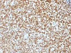
Biotium NKX2.2 (Neuroendocrine & Ewing s Sarcoma Marker) (NX2/1422R), CF488A conjugate, 0.1mg/mL
Expression of NKX2.2 has been found in neuroendocrine tumors of the gut, making it a potential marker for the study of gastrointestinal neuroendocrine tumors. More recently, NKX2.2 protein was identified as a target of EWS-FLI-1, the fusion protein specific to Ewing sarcoma, and was shown to be differentially upregulated in Ewing sarcoma on the basis of array-based gene expression analysis. It acts as a valuable marker for Ewing sarcoma, with a sensitivity of 93% and a specificity of 89%, and aids in the differential diagnosis of small round cell tumors.Primary antibodies are available purified, or with a selection of fluorescent CF Dyes and other labels. CF Dyes offer exceptional brightness and photostability. Note: Conjugates of blue fluorescent dyes like CF405S and CF405M are not recommended for detecting low abundance targets, because blue dyes have lower fluorescence and can give higher non-specific background than other dye colors.
Biotium Cytokeratin 8/18(C-43 + DC10), CF740 conjugate, 0.1mg/mL
Cytokeratin 8 (CK8) belongs to the type II (or B or basic) subfamily of high molecular weight cytokeratins and exists in combination with cytokeratin 18 (CK18). This MAb cocktail recognizes all simple epithelia including glandular epithelium, for example thyroid, female breast, gastrointestinal tract, respiratory tract, and urogenital tract including transitional epithelium. All adenocarcinomas and most squamous carcinomas are positive but keratinizing squamous carcinomas are usually negative. This antibody is useful in demonstrating the presence of Paget cells; there is very little keratin 18 in the normal epidermis so only Paget cells are stained.Immunohistochemical staining with this MAb is indistinguishable from that obtained with monoclonal antibody 5D3.Primary antibodies are available purified, or with a selection of fluorescent CF Dyes and other labels. CF Dyes offer exceptional brightness and photostability. Note: Conjugates of blue fluorescent dyes like CF405S an
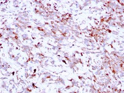
Biotium Cathepsin K (Marker of Tumor Invasiveness)(CTSK/2791), CF488A conjugate, 0.1mg/mL
Cathepsin K is a lysosomal and secreted cysteine proteinase involved in bone remodeling and resorption. This protein, which is a member of the peptidase C1 protein family, is predominantly expressed in osteoclasts. However, the encoded protein is also expressed in a significant fraction of human breast cancers, where it could contribute to tumor invasiveness. Mutations in this gene are the cause of pycnodysostosis, an autosomal recessive disease characterized by osteosclerosis and short stature. Primary antibodies are available purified, or with a selection of fluorescent CF Dyes and other labels. CF Dyes offer exceptional brightness and photostability. Note: Conjugates of blue fluorescent dyes like CF405S and CF405M are not recommended for detecting low abundance targets, because blue dyes have lower fluorescence and can give higher non-specific background than other dye colors.
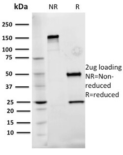
Biotium Fibronectin(Fn-3), CF488A conjugate, 0.1mg/mL
Fibronectins are disulfide-linked, dimeric glycoproteins of ~440 kDa. They possess at least four binding sites for collagen, glycosaminoglycans, transglutaminase, and a cell surface receptor. Epitope of this MAb is located in the 2nd-3rd type-III repeats of fibronectin. Fibronectins are extracellular matrix glycoproteins that are essential for embryonic development. Fibronectins are also involved in cell adhesion, tissue organization, and wound healing. Fibronectins are present in basement membranes, interstitial connective tissue matrix, and blood. Cellular fibronectin is widely distributed in the stroma of many malignant tumors. This MAb reacts with human cellular fibronectin, but not plasma fibronectin.Primary antibodies are available purified, or with a selection of fluorescent CF Dyes and other labels. CF Dyes offer exceptional brightness and photostability. Note: Conjugates of blue fluorescent dyes like CF405S and CF405M are not recommended for detecting low ab
Biotium Chromogranin A(CHGA/777), CF740 conjugate, 0.1mg/mL
Chromogranin A is present in neuroendocrine cells throughout the body, including the neuroendocrine cells of the large and small intestine, adrenal medulla and pancreatic islets. It is an excellent marker for carcinoid tumors, pheochromocytomas, paragangliomas, and other neuroendocrine tumors. Co-expression of chromogranin A and neuron specific enolase (NSE) is common in neuroendocrine neoplasms. Reportedly, co-expression of certain keratins and chromogranin indicates neuroendocrine lineage. The presence of strong anti-chromogranin staining and absence of anti-keratin staining should raise the possibility of paraganglioma. The co-expression of chromogranin and NSE is typical of neuroendocrine neoplasms. Most pituitary adenomas and prolactinomas readily express chromogranin.Primary antibodies are available purified, or with a selection of fluorescent CF Dyes and other labels. CF Dyes offer exceptional brightness and photostability. Note: Conjugates of blue fluorescent dyes
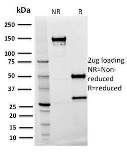
Biotium Spastin(Sp 3G11-1), CF740 conjugate, 0.1mg/mL
The AAA protein family members share an ATPase domain and have roles in various cellular processes including intracellular motility, membrane trafficking, proteolysis, protein folding and organelle biogenesis. Spastin, a member of the AAA protein family, is a 616 amino acid protein and is involved in the function or assembly of nuclear protein complexes. The Spastin protein is expressed ubiquitously and localizes to the nucleus and the cytoplasm, where it may also be involved in microtubule dynamics. Mutations in the Spastin gene (SPAST, SPG4) cause the most common form of spastic paraplegia 4, an autosomal dominant form of hereditary spastic paraplegia (HSP). HSPs comprise a group of inherited neurological disorders characterized by spastic lower extremity weakness due to a length-dependent, retrograde axonopathy of corticospinal motor neurons. SPAST-specific mutations account for approximately 40% of all autosomal dominant HSPs. Primary antibodies are available purified, or
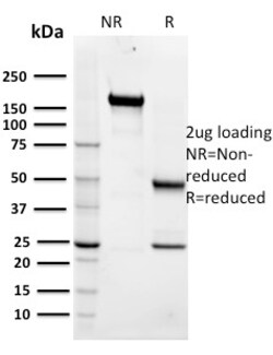
Biotium Maltose Binding Protein / MBP-probe(R29.6), CF568 conjugate, 0.1mg/mL
Plasmid vectors for the expression of coding regions of eukaryotic genes in bacterial, insect and mammalian hosts are in common usage; such expression vectors frequently encode hybrid fusion proteins consisting in part of prokaryotic and in part, eukaryotic specified proteins. One such system utilizes maltose binding protein (MBP), the 370 amino acid product of the E. coli mal E gene. Plasmid vectors have been constructed utilizing the MBP domain that allow the synthesis of high levels of MBP-fusion proteins that can be Purified in a one step procedure by affinity chromatography crosslinked amylose resin. Once bound to amylose, the MBP protein can then be separated from the target protein by cleavage by coagulation factor Xa at a specific four residue site. Alternatively, the intact fusion protein can be specifically eluted from the resin by the addition of excess free maltose. Subsequent to elution, MBP fusion protein can be visualized either by western blot analysis or immu
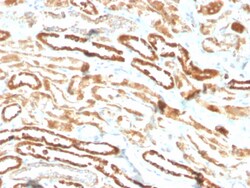
Biotium Erythropoietin (EPO) (Marker of Placentation Disorders)(EPO/3793R), Biotin conjugate, 0.1mg/mL
Recognizes a protein of about 37 kDa, which is identified as Erythropoietin (EPO). Erythropoietin is a secreted, glycosylated cytokine hormone composed of four alpha helical bundles. It is the primary factor responsible for regulating erythropoiesis during steady-state conditions and in response to blood loss and hemorrhage in the adult organism. Erythropoietin is synthesized by the kidney and stimulates the proliferation and maturation of bone marrow erythroid precursor cells. The protein is found in the plasma and regulates red cell production by promoting erythroid differentiation and initiating hemoglobin synthesis. Primary antibodies are available purified, or with a selection of fluorescent CF Dyes and other labels. CF Dyes offer exceptional brightness and photostability. Note: Conjugates of blue fluorescent dyes like CF405S and CF405M are not recommended for detecting low abundance targets, because blue dyes have lower fluorescence and can give higher non-specific
Biotium GFAP (Astrocyte & Neural Stem Cell Marker) (ASTRO/1974R), CF488A conjugate, 0.1mg/mL
This MAb recognizes a protein of ~50 kDa which is identified as Glial Fibrillary Acidic Protein (GFAP). It shows no cross-reaction with other intermediate filament proteins. GFAP is specifically found in astroglia. GFAP is a very popular marker for localizing benign astrocyte and neoplastic cells of glial origin in the central nervous system. Antibody to GFAP is useful in differentiating primary gliomas from metastatic lesions in the brain and for documenting astrocytic differentiation in tumors outside the CNS.Primary antibodies are available purified, or with a selection of fluorescent CF Dyes and other labels. CF Dyes offer exceptional brightness and photostability. Note: Conjugates of blue fluorescent dyes like CF405S and CF405M are not recommended for detecting low abundance targets, because blue dyes have lower fluorescence and can give higher non-specific background than other dye colors.
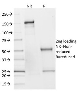
Biotium SDHB (Succinate Dehydrogenase B) (Pheochromocytoma Marker)(SDHB/2382), CF568 conjugate, 0.1mg/mL
Succinate dehydrogenase (SDH) is Complex II in the mitochondria, vital for mitochondrial electron transport, as well as Krebs cycle function. Four subunits comprise the SDH protein complex: a flavochrome subunit (SDHA), an iron-sulfur protein (SDHB) and two membrane-bound subunits (SDHC and SDHD) anchored to the inner mitochondrial membrane. The SDH complex functions as a tumor suppressor. Loss of any subunit proteins lead to destabilization of the complex and tumor formation. Antibody to SDHB is helpful in the identification of phaeochromocytomas, paragangliomas and GIST. Primary antibodies are available purified, or with a selection of fluorescent CF Dyes and other labels. CF Dyes offer exceptional brightness and photostability. Note: Conjugates of blue fluorescent dyes like CF405S and CF405M are not recommended for detecting low abundance targets, because blue dyes have lower fluorescence and can give higher non-specific background than other dye colors.
Biotium p21 / WAF1(CIP1/823), CF740 conjugate, 0.1mg/mL
This MAb recognizes a 21 kDa protein, identified as the p21WAF1 tumor suppressor protein. This MAb is highly specific to p21 and shows no cross-reaction with other closely related mitotic inhibitors. p21WAF1 is a specific inhibitor of cdk s and a tumor suppressor involved in the pathogenesis of a variety of malignancies. The expression of this gene acts as an inhibitor of the cell cycle during G1 phase and is tightly controlled by the tumor suppressor protein p53. Its expression is induced by the wild type, but not mutant, p53 suppressor protein. Normal cells generally display a rather intense nuclear p21 expression. Loss of p21 expression has been reported in many carcinomas (gastric carcinoma, non-small cell lung carcinoma, thyroid carcinoma).Primary antibodies are available purified, or with a selection of fluorescent CF Dyes and other labels. CF Dyes offer exceptional brightness and photostability. Note: Conjugates of blue fluorescent dyes like CF405S and CF405M are